
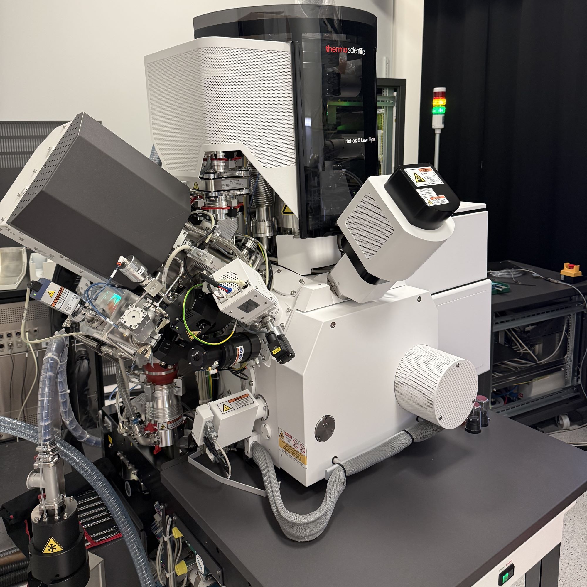
It integrates high-resolution SEM, plasma FIB, and femtosecond laser ablation with Oxford EDS/EBSD, TOFWERK ToF-SIMS, and cryogenic capabilities for rapid 3D characterization, site-specific milling, TEM sample preparation, and multimodal imaging and analysis from the millimeter to nanometer scale.
This instrument was acquired through support from the U.S. National Science Foundation under MRI Grant No. 2511067.
| Column | Elstar electron column with UC+ technology |
| Source | Schottky field emitter |
| Objective lens | Dual-mode magnetic immersion / field-free |
| Accelerating voltage | 350 V – 30 kV (20 V – 30 kV with beam deceleration) |
| Beam current | 0.8 pA – 100 nA |
| Resolution | 0.6 nm at 2–15 keV · 0.7 nm at 1 keV · 1.0 nm at 500 eV · 1.1 nm at 300 eV |
| Detectors | TLD (SE/BSE) · ICD (low-loss BSE) · ETD (SE) |
| Ion species | Xe, Ar, O2, N2 — selectable |
| Accelerating voltage | 500 V – 30 kV |
| Beam current | 1 pA – 2.5 µA in 14 steps at 30 kV |
| Resolution | < 20 nm at 30 kV (Xe) |
| Integration | In-chamber, coincident point shared with SEM and FIB |
| Wavelength | 1030 nm (IR) · 515 nm (green) |
| Pulse duration | < 300 fs |
| Repetition rate | 1 kHz – 1 MHz |
| EDS | Oxford Ultim Max, 100 mm² SDD |
| EBSD | Oxford Symmetry 3 |
| SIMS | ToF-SIMS C2 — elemental and isotopic 3D imaging |
| Stage | 5-axis · X-Y 150 mm · tilt −38° to +53.8° · 360° rotation · 100 nm minimum step |
| Vacuum | Oil-free with integrated plasma cleaner; cryo modules available |
| Lamella transfer | EasyLift nanomanipulator |
| Automated prep | AutoTEM 5 for STEM/TEM specimens |
| Deposition | GIS — W, Pt |
| 3D workflows | Auto Slice & View for automated serial-section datasets |
| Software | AutoScript (Python) · MAPS correlative · Avizo |
Complete the Helios training session with staff before independent use.
Science Education and Research Center (SERC)
Room 008
Publications using EM Lab data must acknowledge the facility.
See here.