Archives

All posts for the month October, 2016

Patient profile

  • 14 year old male
  • Spastic diplegic Cerebral Palsy
  • GMFCS Level II
  • History of tendon lengthening
  • Forearm crutches for community distances
  • Has not been attending PT regularly
  • Cognitively intact and motivated

Patient goals

  • Participate in physical education class
  • Play basketball without requiring rest breaks

Example of GMFCS Levels:

PICO

What are the effects of functional strength training versus typical physical therapy on walking ability in a pediatric population (<22 years of age) with spastic cerebral palsy?

Search Strategy 

100 articles identified

in: PubMed, CINAHL, OVID- Medline

16 duplicates removed

84 records screened

74 not relevant

10 full text screened

4 full text excluded

6 included

Inclusion Criteria

  • Pediatric Population (<22 years old) with a diagnosis of spastic cerebral palsy who are also ambulatory
  • Interventions that include strength or resistance training of lower extremities
  • Assessment of at least one of the following: 6 minute walk test, TUG, 20 meter walk test, GMFM D/E

Exclusion Criteria

  • Patient’s who had undergone botox injections within the past 3 months
  • Surgery in the past 6 months
  • Non-ambulatory patients
  • Adult population
  • Upper extremity training
  • Inability to follow direction or cognitive/behavioral diagnosis

Results 


Limitations

  • Small sample sizes, underpowered
  • Cross-contamination of intervention
  • Undefined control interventions; “typical PT”
  • Lack of randomized controlled trials
  • Lack of blinding
  • Recruitment of convenience

Clinical Bottom Line

There is conflicting and limited, low quality, evidence that suggests that strength training does not improve walking ability in pediatric populations with spastic cerebral palsy with diagnoses that fall into GMFCS Levels I, II, & III.

  • 2 studies were randomized controlled trials, both looking specifically at strength training, and lacking defined controls. Neither of these studies found significant improvement in any measures of walking ability, such as the TUG or GMFM D/E.
  • 3 studies, all lower level evidence; case studies or quasi-experimental studies, found evidence that strength training improved measures of walking ability, but did not have control groups and included other intervention strategies within their therapeutic sessions, such as dynamic balance or gait training.
  • The final study found that strength training and treadmill training both led to clinically detectable improved walking ability.

Application to Case

Strength training should be included in a standard physical therapy plan of care if it aligns with patient-specific goals or to specifically increase strength. Interventions that focus on gait training and walking endurance may be more beneficialwhen implemented as primary interventions to improve walking ability

Strength training does not appear to cause any negative effects to patients with spastic CP, although range of motion and spasticity should be monitored if strength training regimens are implemented.

References

Auld ML, Johnston LM. “Strong and steady”: a community-based strength and balance exercise group for children with cerebral palsy. Disabil Rehabil. 2014;36(24):2065-71.

Aviram R, Harries N, Namourah I, Amro A, Bar-haim S. Effects of a group circuit progressive resistance training program compared with a treadmill training program for adolescents with cerebral palsy. Dev Neurorehabil. 2016:1-8.

Aye T, Thein S, Hlaing T. Effects of strength training program on hip extensors and knee extensors strength of lower limb in children with spastic diplegic cerebral palsy. J Phys Ther Sci. 2016;28(2):671-6.

Dos Santos AN, da Costa CS, Golineleo MT, Rocha NA. Functional strength training in child with cerebral palsy GMFCS IV: case report. Dev Neurorehabil. 2013 Oct;16(5):308-14. doi: 10.3109/17518423.2012.731085. Epub 2013 Apr 25. PubMed PMID: 23617884.

Scholtes VA, Becher JG, Janssen-Potten YJ, Dekkers H, Smallenbroek L, Dallmeijer AJ. Effectiveness of functional progressive resistance exercise training on walking ability in children with cerebral palsy: a randomized controlled trial. Res Dev Disabil. 2012 Jan-Feb;33(1):181-8. doi: 10.1016/j.ridd.2011.08.026. Epub 2011 Oct 11. PubMed PMID: 22093663.

Taylor NF, Dodd KJ, Baker RJ, Willoughby K, Thomason P, Graham HK. Progressive resistance training and mobility-related function in young people with cerebral palsy: a randomized controlled trial. Dev Med Child Neurol. 2013 Sep;55(9):806-12. doi: 10.1111/dmcn.12190. Epub 2013 Jun 22


Background Information:

dps-illustration-text_0

  • 4% of spinal cord injuries (SCI) lead to chronic mechanical ventilation
  • Mechanical ventilation, currently the gold standard for respiratory dependent patients, has many potential complications
  • Serious complications include:
    • Pneumonia, the leading cause of decreased life expectancy amongst this population
    • Respiratory Infection
  • Via transcutaneous electrical stimulation, the patency of the phrenic nerve can be determined preoperatively
  • If intact, these patients may be candidates for phrenic nerve pacing
  • Phrenic nerve pacers can be laparoscopically placed, including internal electrodes and an external transmitter

Clinical Scenario:

A 36 year old male sustained a C3 ASIA B SCI 1 week ago He is currently in the ICU being mechanically ventilated with no significant improvement. Prior to his injury, he was living at home with his wife and two young children and working as an investment banker. Physical examination findings include diminished sensation throughout thoracic region and no motor function below level of injury. Patient goals are to return to home and work independently with focus on resuming his role as a father.

PICO Question:

Does diaphragmatic pacing improve respiratory independence and QOL in individuals with spinal cord injury as compared to mechanical ventilation.

Search Strategy:

21 articles identified using PubMed, CINAHL and Cochrane.

Variations of Terms Used: “spinal cord injury”, “diaphragm pacing”, “respiratory”, “phrenic nerve”

Inclusion Criteria: Diagnosis of cervical spinal cord injury, ventilator dependence, English studies, published 2005-present

Exclusion Criteria: Diagnosis of  Amyotrophic Lateral Sclerosis, phrenic nerve lesions

Results:

Author

Year

Country

Patient Population Study Type and Level Of Evidence Outcomes Key Results
Tedde ML, et. al. (2012)

Brazil

Ventilator dependent quadriplegics post cervical trauma Clinical Trial

Level 3 Evidence

D&B: 12/27

Independence from MV3 •N=5

•At 6 month f/u:

•3 patients continuous DPS4

•1 patients 6 hours/day

•1 patient unable to sustain

Romero FJ, et. al. (2012)

Spain

Ventilator Dependent High-Cervical SCI patients Retrospective Review

Level 3 Evidence

D&B: 17/27

SF-36 •Social Functioning:

•PNP: 7.67 ± 1.80

•MV Group: 5.67 ± 1.17

•P value <.001

•No significant change in physical functioning, general health, role limitations, physical pain, vitality

Poluszny JA, et. al. (2013)

United States

Ventilator dependent SCI patients with intact phrenic nerves Retrospective Cohort Study

Level 3 Evidence

D&B: 16/27

Independence from MV3 •N=22

•18 participants continuous DPS4

•2 participants: 4 and 12 hours/day

•1 patient unable to sustain

•1 patient with prolonging life measures withdrawn

Hirschfeld S, et. al. (2008)

Germany

SCI patients with induced respiratory device dependency Prospective Cohort Study

Level 3 Evidence

D&B: 20/27

Patient and Physician reported QOL •Unanimous patient reported improved self confidence with PNP over MV
Onders RP, et. al. (2007)

United States

Ventilator dependent tetraplegics with a C2 injury occurring in childhood Prospective Cohort Review

Level 3 Evidence

D&B: 9/27

Independence from MV3, Patient reported QOL •N=10

•4 patients continuous DPS4

•4 patients day time only

•2 unable to sustain

•Unanimous patient reported preferences for DPS4, improvements in mobility and ease of integration into society

Limitations:

  • Lacking validated outcome measures
  • Small Sample Sizes
  • Procedures for implantation and weaning were variable
  • Lacking long term follow up
  • No control groups
  • Lack of homogeneity amongst groups
    • Age
    • Time on MV
    • Level of injury
    • Severity of Injury

Clinical Bottom Line:

There is low-level evidence that suggests phrenic nerve pacing is a safe and effective alternative treatment for ventilator dependent, high-level, cervical spinal cord injury patients. Additionally, this evidence supports decreased complications common with mechanical ventilation and improved resocialization, mobility, independence, and overall quality of life.

Application:

Phrenic nerve pacing has been shown to improve independence and quality of life. Due to my patient’s goals of returning to his work and family responsibilities, and the lack of any serious adverse events reported with this treatment, phrenic nerve pacing may be an appropriate alternative. After transcutaneous nerve testing determines the patency of his phrenic nerve, offering this as a potential alternative treatment to chronic mechanical ventilation allows my patient to be an active participant in his plan of care.

Acknowledgements:

Thank you to Dr. Richard Lauer for your assistance along the way.

References:

  1. Posluszny JA, Onders R, Kewin AJ, Weinstein MS, Stein DM, Knight J, et al. Multicenter review of diaphragm pacing in spinal cord injury: Successful not only in weaning from ventilators but also in bridging to independent respiration. J Trauma Acute Care Surg. 2014;76(2):303-9. Doi:10.1097/TA.0000000000000112
  2. Romero FJ, Gabarrutta C, Garcia-Forcada A, et al. Long-term evaluation of phrenic nerve pacing for respiratory failure due to high cervical spinal cord injury. Spinal Cord. 2012; 50(12): 895-8. Doi: 10.1038/sc.2012.74
  3. Tedde ML, Filho PV, Hajjar LA, Almeida JP, Flora GF, et. al. Diaphragmatic pacing stimulation in spinal cord injury: anesthetic and perioperative management. Clinics. 2012. 67(11). Doi: 10.6061
  4. Hirschfeld S, Exner G, Luukaala T, Baer GA. Mechanical ventilation or phrenic nerve stimulation for treatment of spinal cord injury-induced respiratory insufficiency. Spinal Cord. 2008; 46(11): 738-42. Doi: 10.1038/sc.2008.43
  5. Onder RP, Elmo MJ, Ignagni AR. Diaphragm Pacing Stimulation System for Tetraplegia in Individuals Injured During Childhood or Adolescence. J Spinal Cord Med. 2007; 30(1):25-29.

With further questions please contact Rebecca Maidansky at rmaidansky@temple.edu

Background:

It is well documented that survivors of stroke are at an increased risk of falls relative to age-matched controls.1,2 Ability to take a reactive step is paramount in recovery from an unexpected loss of balance.3,4 Therefore impaired reactive stepping has been proposed as one important component of the multivariate nature of falls following a stroke. One value that has been the topic of discussion in reactive stepping literature is the TFO, or time to foot off. This measure represents the time between the release of external support of an individual leaning their center of mass (anteriorly) outside of their base of support, to the time in which their body weight is no longer supported by the limb in which they choose to step. Characteristics of impaired reactive stepping have been shown to correlate with falls in elderly populations.2

img_2087

Search Strategy:

Pub Med:  Search Terms: (((stepping) OR reactive stepping) AND poststroke) OR reactive stepping[Title/Abstract]

CINAHL: Boolean/phrase: reactive stepping OR stepping AND poststroke

WOS: Search strategy : TITLE: (reactive stepping) AND TOPIC: (stroke)

Inclusion Criteria:

-Participants must be above the age of eighteen and had their neurologic incident at least two weeks prior to enlisting in the study in order to be considered ‘chronic.’

-The methodology of how the researchers obtained values related to perturbation or reactive response must be clearly explained within the article.

-At least one outcome related to falls in the stoke population must have been collected.

Exclusion Criteria

-Articles which only examined metrics on gait or focused on outcomes related to gait.

-Level V evidence

uno

dos

tres


Rationale for data collection:

A lack of association between reactive stepping impairment and utilized clinical measures (BBS, gait velocity, STRATIFY falls risk tool) has been identified. This suggests a limitation may exist when drawing inferences with these measures to predict likelihood of falls. Therefore we may be missing a critical evaluative component in this patient population, which could also help us streamline our intervention. In addition, falls render increased healthcare utilization and diminished quality of life of affected individuals.

Methods:

-Setting and participants:

Leahy lab at the University of the Sciences in Philadelphia. Subjects (n=6) recruited as part of a larger study at the university lab investigating moderate intensity walking in people with stroke. Subjects recruited from local physical therapy clinics, stroke support groups, and individuals who have participated in research and classroom activities previously at the USciences Department of Physical Therapy. Healthy controls (n=5) are healthcare students recruited by convenience.

Inclusion:

-People over the age of 18 years with one or more strokes at least 3 months previously

-Ambulatory without physical assistance of other person (orthotic devices, canes or walkers will be allowed)

-Able to follow instructions and able to communicate with investigators as assessed by specific NIH Stroke Scale questions.

Exclusion:

-Bone or joint problems that limit the ability to walk

-Resting hear rate outside of the range of 40-100 beats per minute

-Resting blood pressure outside of the range of 90/60 to 170/90 mm Hg

-Chest pain or shortness of breath with exertion

-Score of >1 on question 1b and >0 on question 1c on the NIH Stroke Scale inability to communicate with investigators.

Demographics:

Control group:

quatro

Affected group:

cinco


Upon entering clinic individuals were oriented to the purposed of the study, screened to be deemed fit for inclusion, and consented for participation. Then the following outcomes were collected: TUG, TUGmanual, TUGcognitive, 6MWT, 4 square step test (modified if unable), gait velocity-preferred, and gait velocity-fast.

They then underwent the reactive stepping assessment. They underwent five trials where they were able to step with whatever limb they desired. Following this five trials were done while blocking the preferred limb in an attempt to elicit a step with the non-preferred limb.

The method for each trial is as follows:

  1. Participant is to stand statically in their preferred foot position inside of a body weight support system designed to catch them if an attempt to regain balance is failed.
  2. Patient will be positioned on a GaitRite system. (Collecting data through ProtoKinetics Movement analysis software)
  3. A gait belt is then locked into a closed loop around the patient by tester #1 and placed outside of the subject’s shoulders, utilizing the acromion bilaterally as the landmark of standardization.
  4. A Lafayette Manual Muscle Testing myometer is then placed inside of the gait belt (with the screen visible to tester #1) so as to garner the force paced upon it when the gait belt pulls forward, as described in step #6.
  5. Tester #1 checks to make sure both feet are in alignment before having subject lean.
  6. As tester #1 holds the myometer statically in place, the individual is instructed to progressively lean forward into the gait belt until 8-10% of their precalculated bodyweight is displayed on the readout. The subject is then instructed to hold this position as best they can.
  7. The subject is then released 2-10 seconds after they are instructed to hold their ‘forward leaning’ position with the desired % body weight. At this time the subject is release by tester #1, inducing a reactive stepping response. If at any point the subject falls outside of the 8-10% range of their bodyweight, the process will be started over again from step #4.

Data Extraction

Extracting the TFO value was done utilizing the PKMAS software system for the GaitRite mat. In the photo below the box labeled ‘temporal 4’ gives the indication of when the subject was released through the sharp increase in slope of the center of pressure (red) reading. This time is noted, and the time at which pressure is no longer picked up by the stepping limb is noted. The time between these two values is noted. The GaitRite mat has a sampling rate of 120 Hertz, therefore frames are captured every 8 milliseconds.

tfo-ex2

Statistical analysis:

Nonparametric

Independent-samples Mann-Whitney U test

-To identify between-group differences in TFO and outcome measures

Spearman rank correlation coefficient (Spearman’s rho)

-To identify correlation between TFO and outcome measures

Wilcoxon signed-ranks test

-To identify between-limb differences in stroke and control group


seis

seite

Results:

**Red indicates statistical significance

Wilcoxon Signed ranks test

-No significant difference between TFOu and TFOr in either group.

M-WU:

ocho

nueve

Limitations:

-Inherent limitation in methodology (e.g. handheld myometer use). Non age-matched controls from sample of convenience. Small sample size limits generalizability and ability to establish validity.

Suggestions for future research:

Larger sample sizes. Research individuals who have experienced a stroke compared to age-matched cohort. Correlate TFO values with fall rates in the community. Explore utility of this measure as an outcome and how it responds to interventions. Cost-effective methods of collecting value in outpatient realm.

Citations

1.Maki B, Edmonstone M, Perry S, Heung E, Quant S, McIlroy W. Control of rapid limb movements for balance recovery: do age-related changes predict falls risk? Control of Posture and Gait. Maastricht, the Netherlands: International Society for Postural and Gait Research; 2001: 126-129.

2.Hilliard MJ, Martinez KM, Janssen I, et al. Lateral balance factors predict future falls in community-living older adults. Arch Phys Med Rehabil. 2008;89: 1708-1713

3.Maki BE, McIlroy WE. The role of limb movement in maintaining upright stance: the “change-in-support” strategy. Phys Ther. 1997;77: 488-507.

4.Maki BE, McIlroy WE. Control of rapid limb movements for balance recovery: age-related changes and implications for fall prevention. Age Ageing. 2006;35(suppl 2): ii12-ii18.

5 articles from search:

  1. Mansfield A, Inness EL, Wong JS, Fraser JE, McIlroy WE. Is impaired control of reactive stepping related to falls during inpatient stroke rehabilitation? Neurorehabil Neural Repair. 2013;27:526-33.
  2. Inness EL, Mansfield A, Lakhani B, Bayley M, McIlroy W. Impaired reactive stepping among patients ready for discharge from inpatient stroke rehabilitation. Phys Ther. 2014;94:1755-64.
  3. Martinez KM, Mille M, Zhang Y, Rogers MW. Stepping in persons poststroke: comparison of voluntary and pertubation-induced responses.  Arch Phys Med Rehabil. 2013; 94:2425-32.
  4. Inness EL, Mansfield A, Bayley M, McIlroy WE. Reactive stepping after stroke: determinants of time to foot off in the paretic and nonparetic limb. Journal of Neurologic Physical Therapy. 2016; 40:196-202.
  5. Lakhani B, Mansfield A, Inness E, McIlroy WE. Compensatory stepping responses in individuals with stroke: a pilot study. Physiotherapy Theory and Practice. 2011; 27(4): 299-309.


Background Information:

  • An estimated 3.8 million people sustain a concussion every year1,4
  • 10% – 33% of individuals with mild traumatic brain injury experience symptoms months to years later1
  • Post-concussion syndrome (PCS) is defined as persistence of > 3 symptoms 7-10 days after injury5: Headache, Dizziness, Fatigue, Irritability, Insomnia, Concentration Difficulty, Memory Difficulty, Stress or emotion intolerance
  • One cause of PCS may be altered autonomic function and impaired autoregulation and distribution of cerebral blood flow

Clinical Case:

  • 15 y.o. female student s/p MVA 1 month ago hitting head on dash board
  • ER visit diagnosed mTBI –> prescribed “rest”
  • S/s:  headaches, fatigue, worsens with reading, noisy family, screen time
  • Goals: asymptomatic school day, return to tennis, recreational running
  • PT: Vestibular, postural, and cervicogenic exercises initiated

PICO: Is aerobic exercise beneficial in individuals with prolonged symptoms after mTBI?


Search Strategy: 1,247 articles identified in PubMed, CINAHL, Academic Search Complete

  • Terms: (concussion OR post-concussion syndrome OR mild traumatic brain injury) AND (exercise OR rehabilitation OR treatment). English language, peer reviewed, humans, 10 years.
  • Inclusion: subjects sustained mild TBI/concussion by means of sport, MVA, or other,  experiencing post-concussive symptoms > 2 weeks, exercise intervention defined, post-concussive related outcome measures (symptoms, fatigue, QOL, function)
  • Exclusion: acute phase mTBIs, moderate to severe TBI’s, non interventional studies

Results:

results-chart

aPost-concussive symptoms > 4 weeks aa > 6 weeks, bBalke Treadmill test, BDI: Beck Depression Inventory, PCSI: Post Concussion Symptom Inventory, PCS: Post Concussion Scale, SE: statistically significant effects listed (p <0.05)

Limitations and suggestions:

  • “Active” control groups1,3, Lack of control group2,3,5
  • Small sample size1-5
  • Multiple treatment interference2
  • Inadequate follow up1-3,5
  • Lacking consensus on intensity and duration
  • Optimal timing of intervention unclear
  • Higher quality research suggested to directly compare aerobic exercise with other interventions such as dual tasking, cervicogenic, vestibular exercises, and non participants.

Clinical Bottom Line:

There is moderate to low level evidence to support the use of aerobic exercise in patients with prolonged symptoms after sustaining a mild traumatic brain injury.  This particular literature review offers recent interventional case studies and a randomized clinical trial in which aerobic exercise is the primary intervention and assessment tool used to prescribe a customized, progressive, aerobic rehabilitation program whether on a treadmill or stationary bike. Each study reported a statistically significant change in some aspects of post-concussive symptoms such as return to full daily functioning, ability to exercise maximally without symptom exacerbation, and decrease in post-concussion symptom specific scales. No adverse events or negative impacts were reported in the studies from engaging sub-symptom exacerbation aerobic exercise in patients still experiencing prolonged symptoms after mTBI, which had previously been thought to be deleterious until research as such as been published in the recent decades.

It is proposed that exercise assessment and aerobic exercise training may reduce concussion-related physiological dysfunction by restoring autonomic balance and by improving autoregulation of cerebral blood flow. Controlled progressive aerobic exercise treatment may help to restore normal CBF regulation by conditioning the brain to gradually adapt to repetitive mild elevations of systolic blood pressure.3-5


Application:

  • Obtain baseline (use of BALKE, BUFFALO CONCUSSION TREADMILL TEST, or Bike Test described in Kurowski 2016)
    • Treadmill or bike
      • Stationary bike program indicated if excess head movements are provoking on treadmill
    • Monitor vitals
      • HR, BP, RPE, & symptoms
    • Progress intensity until exacerbation
  • Prescribe aerobic exercise program
    • Intensity: 80% of symptomatic duration OR symptomatic heart rate/RPE if patient able to self monitor
    • Frequency: 5 days a week, 4 to 6 weeks
    • Monitor symptom trends and modify program accordingly weekly
      • progress it!
  • Consider multi-modal treatment
    • Though aerobic exercise seems beneficial in ameliorating prolonged symptoms, consider challenging your patient in other modes such as dual tasking, vestibular, balance, and sport specific exercises alongside your aerobic exercise treatment (see Gagnon 2016) for they may be symptomatic in some realms but not others.

Acknowledgements:

Anne Galgon, PT, PhD, NCS

Contact: Jackie Pedersen, Temple Class of 2017. jacqueline.pedersen@temple.edu

References:

1. Kurowski BG, Hugentobler J, Quatman-Yates C, Taylor J, Gubanich PJ, Altaye M, Wade SL. Aerobic Exercise for Adolescents With Prolonged Symptoms After Mild Traumatic Brain Injury: An Exploratory Randomized Clinical Trial.  J Head Trauma Rehabil. 2016 Apr 26. [Epub ahead of print] PubMed PMID: 27120294.

2.Gagnon I, Grilli L, Friedman D, Iverson GL. A pilot study of active rehabilitation for adolescents who are slow to recover from sport-related concussion. Scand J Med Sci Sports. 2016;26(3):299-306

3.Leddy JJ, Cox JL, Baker JG, Wack DS, Pendergast DR, Zivadinov R, Willer B. Exercise treatment for postconcussion syndrome: a pilot study of changes in functional magnetic resonance imaging activation, physiology, and symptoms. J Head Trauma Rehabil. 2013 Jul-Aug;28(4):241-9

4. Baker JG, Freitas MS, Leddy JJ, Kozlowski KF, Willer BS. Return to full functioning after graded exercise assessment and progressive exercise treatment of postconcussion syndrome. Rehabilitation Research & Practice. 2012:1-7

5.Leddy JJ, Kozlowski K, Donnelly JP, Pendergast DR, Epstein LH, Willer B. A preliminary study of subsymptom threshold exercise training for refractory post-concussion syndrome. Clinical Journal of Sport Medicine. 2010;20(1):21-2


Background:

  • Neck pain is the third most commonly reported type of pain
  • There is a higher prevalence in women than men
  • Chronic neck pain often leads to impaired movement coordination, endurance, and strength


Clinical Case and PICO:

  • 59 y.o. retired female seamstress with insidious onset of neck pain that has lasted over 5 yrs.
  • Current pain level of 4/10 and 8/10 at worst on NPRS| NDI: 30%
  • Denies any numbness, tingling, and headaches
  • Relief with lying down and NSAIDs
  • Family physician advised that general exercise would ↓ pain
  • Primary Impairments: ↓neck ROM (flexion, b/l rotation), ↓ endurance (per CCFT)
  • Functional Limitations: reading, crossing the street, care-taking grandson

PICO: For individuals with chronic neck pain, is general exercise as effective for reducing pain and improving function as specific exercise targeting deep neck flexors?


picture2

Figure 1: Deep Cervical Flexors – Longus Capitis and Longus Colli


Search Strategy:

  • 59 articles identified
  • via PubMed, CINAHL, and PEDro
  • 9 duplicates removed
  • 50 articles screened
  • 42 articles not relevant
  • 8 full-text articles screened
  • 3 articles excluded
  • 5 articles included


Key Results:

Author, Date, Country Patient Population Study Type, Evidence Outcomes Key Results
O’Leary, 2011, Australia N = 60

Pain > 6 mos., score btw. 10/50 – 15/50 on NDI

RCT

Level II

Pain (VAS)

Disability (NDI)

Specific exercises targeting endurance, strength, coordination, and ROM improved these respective motor performances with minor carry-over in performance in other motor domains.

Pain: Meaningful from baseline to 10 wks. for coordination group (↓19.2 mm)

Disability: All groups had similar reductions in disability

Ludvigsson, 2015, Sweden N = 216

Pain > 6 mos., > 10/50 on NDI,  > 20/100mm on VAS, post-WAD.

RCT

Level II

Pain (VAS)

Disability (NDI)

Neck-specific group resulted in better outcomes in pain and disability than general exercise group.

Pain: Significant ↓ over time in all three groups

Disability: Significant ↓ from baseline to 6 mos. for neck-specific group.

No improvement for general exercise.

Izquierdo, 2015, Spain N = 28

Pain > 3 mos., < 15/50 on NDI

RCT

Level II

Pain (VAS)

Disability (NDI)

Both CCF and proprioceptive groups  improved in pain and disability as well as positive outcomes for deep neck flexor neuromuscular control.

Pain: Meaningful for current pain from baseline to post 2 mos. (↓3.45mm in CCF and ↓4.00mm in proprioception). Resulted in no current pain.

Disability: Significant from baseline to post 2 mos. (↓3.25pts. for CCF and ↓3.28pts. for proprioception)

Borisut 2013, Thailand N = 100

Pain > 6 mos., > 30/100 mm on VAS

Clinical Trial

Level III

Pain (VAS)

Disability (NDI)

Pain and disability improved for strength-endurance, CCF, and combined group.

Pain: Meaningful from baseline to 12 wks. in combined group (↓44.6mm)

Disability: Meaningful from baseline to 12 wks. in strength-endurance (↓13.51pts.), CCF (↓15.55pts.), combo (↓13.52pts.)


Kim, 2016, South Korea N = 28

Pain > 3 mos., NDI < 15 pts.

Clinical Trial

Level III

Pain (NPRS)

Disability (NDI)

DCF group had better outcomes in pain and disability than general exercise group.

Pain: Meaningful at 8 wks. for DCF (↓3.5pts.)

Disability: Significant at 8 wks. for DCF (↓3.8 pts.)

Legend: CCF = craniocervical flexion  | VAS = Visual Analog Scale | CCFT = craniocervical flexion test

DCF = deep cervical flexor  | NDI = Neck Disability Index | NPRS = Numeric Pain Rating Scale


Clinical Bottom Line:

Upon review of moderate level evidence comparing general exercise to deep neck flexor exercise in treating chronic neck pain, general exercise is not as effective as deep neck flexor exercise but more specifically, the evidence suggests that any neck-specific exercise can improve pain and disability.


Limitations:

  • No long-term follow-up past 6 months
  • Only studied patients with mild neck pain and disability
  • Subjects and therapists were not blinded
  • Impairments were not targeted for patient interventions


Application:

As with most conditions, it is imperative to identify the key impairments related to the individual and to prescribe exercises that target that impairment.  The specific neck exercise that targets the impairment will address the impairment and also improve pain and disability.

Plan of Care –

Patient Education:

– Ergonomic modifications

– Discuss chronic pain

– Reassurance

Manual Therapy:

– Cervical Traction

– Atlantoaxial Joint Mobilization

Therapeutic Exercise:

– Endurance – CCF

– Mobility – cervical rotation and flexion

picture1

Figure 2: Craniocervical Flexion Exercise using objective measurement


Acknowledgements:

Bill Egan, PT, DPT, OCS, FAAOMPT & Temple DPT Class of 2017


References:

1. O’Leary S, Jull G, Kim M, Uthaikhup S, Vicenzino B. Training mode-dependent changes in motor performance in neck pain. Arch Phys Med Rehabil. 2012;93(7):1225-1233. doi: 10.1016/j.apmr.2012.02.018 [doi].

2. Ludvigsson ML, Peterson G, O’Leary S, Dedering A, Peolsson A. The effect of neck-specific exercise with, or without a behavioral approach, on pain, disability, and self-efficacy in chronic whiplash-associated disorders: A randomized clinical trial. Clin J Pain. 2015;31(4):294-303. doi: 10.1097/AJP.0000000000000123 [doi].

3. Gallego Izquierdo T, Pecos-Martin D, Lluch Girbés E, et al. Comparison of cranio-cervical flexion training versus cervical proprioception training in patients with chronic neck pain: A randomized controlled clinical trial. J REHABIL MED (16501977). 2016;48(1):48-55. http://libproxy.temple.edu/login?url=http://search.ebscohost.com/login.aspx?direct=true&db=cin20&AN=112739640&site=ehost-live&scope=site. doi: 10.2340/16501977-2034.

4. Borisut S, Vongsirinavarat M, Vachalathiti R, Sakulsriprasert P. Effects of strength and endurance training of superficial and deep neck muscles on muscle activities and pain levels of females with chronic neck pain. J PHYS THER SCI. 2013;25(9):1157-1162. http://libproxy.temple.edu/login?url=http://search.ebscohost.com/login.aspx?direct=true&db=cin20&AN=104154602&site=ehost-live&scope=site. doi: 10.1589/jpts.25.1157.

5. Kim JY, Kwag KI. Clinical effects of deep cervical flexor muscle activation in patients with chronic neck pain. J Phys Ther Sci. 2016;28(1):269-273. doi: 10.1589/jpts.28.269 [doi].


Contact Information:

georgechau@gmail.com

Background:

  • More than 795,000 people suffer from a cerebral vascular accident (CVA) each year
    • Ischemic strokes account for approximately 87% of all CVA’s
  • Leading cause of disability in the United States
  • CVA’s cost an estimated $34 billion each year
    • Includes medication costs, health care services, and missed work days
  • Repetitive Transcranial Magnetic Stimulation (rTMS) is a device that uses an electromagnet to stimulate small areas of the brain
    • This is achieved by low or high frequency magnetic pulses that send an electric current across the brain tissue
  • Low frequency stimulation has inhibitory effects
    • Transcallosal Disinhibition: By inhibiting the non-lesioned hemisphere the theory is that competition between hemispheres is decreased and it allows for more cortical excitability due to the non-lesioned hemisphere no longer inhibiting the affected hemisphere.
  • High frequency stimulation has excitatory effects

picture1

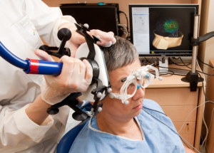
Image source: www.practicalpainmanagement.com

Case Scenario:

  • Hank is a 66 y.o. male diagnosed with a left middle cerebral artery thrombotic infarction 2 weeks ago.  He underwent surgery for recanalization of his left MCA and was placed on anticoagulants. Hank has a past medical history of hypertension, diabetes mellitus II, and peripheral vascular disease.

    Upon exam Hank was A&O x 3, able to follow commands, and provide social history. He is a retired school teacher who enjoys working on antique cars in his free time. Hank lives at home in a 1 story house with his wife and was previously independent with all ADLs and IADLs. He denies any pain but states, “I just can’t really feel my left hand very well.”  Hank is 6’1” and 182 lbs and right hand dominant.


    Hank is very hopeful that PT will “get his arm moving like it used to” so he has the upper extremity strength and hand dexterity to continue working on his cars.

picture2

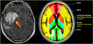
Image Source: www.radiologyassistant.nl

Key Exam Findings:

  • Right upper extremity demonstrates a flexion synergy pattern with AROM
  • Manual muscle tests of the right upper extremity
    • Shoulder flexion 3/5
    • Elbow flexion 2/5
    • Elbow extension 2/5
    • Decreased grip strength
  • Diminished sensation to light touch in right upper extremity throughout C6-T1 dermatomes

PICO Question:

Does repetitive transcranial magnetic stimulation before activity training enhance upper extremity motor recovery in adults s/p CVA?
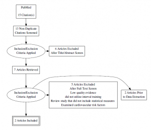
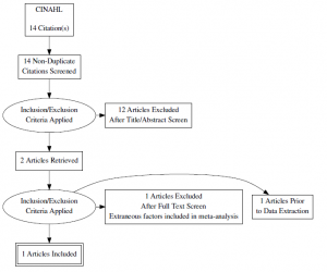
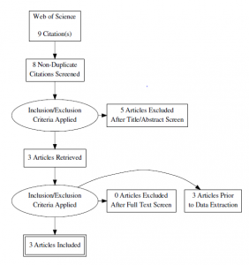
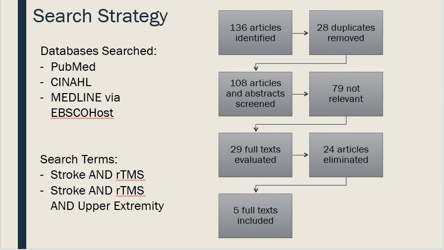
Search Strategy:

The following limits were applied: Published within the past 10 years, human species, English language, Meta-analyses, Systematic Reviews, RCTs

try

Inclusion Criteria, article must include:

  • Adults (>18 years old) who present with upper extremity paresis after stroke
  • At least one intervention arm that consisted of repetitive transcranial magnetic stimulation to either the ipsilesional or contralesional hemisphere
  • At least one intervention arm that consisted of motor/activity training after stimulation
  • Assessment of at least one of the following outcome measures:
    • Fugl- Meyer Assessment
    • Wolf Motor Function Test
    • 9 Hole Peg Test
    • Grip strength
    • Upper limb force production

Exclusion Criteria, article cannot include:

  • Stimulation to areas of the brain other than M1 or the premotor cortex
  • Other non-invasive brain stimulation techniques

Evidence Summary Table:

Author, Date, and Country Patient Population Study Type and Level of Evidence Outcomes Conclusion Study Weaknesses
Hao, 2013

China

Patients of any age or sex after stroke, regardless of the duration of illness or severity of the initial impairment Systematic Review

Level I

PRISMA: 23/27

Motor Outcomes: Jebsen-Taylor Hand Function Test, Pegboard Task, Wolf Motor Function Test, Action Research Arm Test –Use of rTMS for patients with stroke is not recommended until its efficacy is verified in high-quality, large scale RCT’s

Subgroup analysis: Contralesional rTMS displayed a moderate effect size on motor outcomes

Small number of subjects across all articles reviewed with sample sizes ranging from 10 to 123.

Stimulation parameters varied across studies.

No studies included with long term follow-up.

Inclusion criteria for participants was not very strict, for example a study with pediatric patients was included.

Meta-Analysis for motor function was only performed on 4 of 19 studies.


Hsu, 2012

Taiwan

Adults diagnosed with stroke Systematic Review

Level I

PRISMA: 20/27

Wolf Motor Function Test, Nine-Hole Peg Test, Grip Strength  

–rTMS suggests a clinically positive effect on motor recovery in the affected upper limb of patients with stroke

Low frequency rTMS over the contralesional hemisphere being more effective in enhancing upper limb motor recovery based on subgroup analysis

Small number of subjects across all articles reviewed

Many articles reviewed report a wide variety of long term outcomes, if any (from 2 weeks to 1 year),

Outcome measures, experimental designs, inclusion criteria, and rTMS protocols varied between articles.


Etoh, 2013

Japan

Adults with chronic stroke; either first or second unilateral stroke Randomized Double-Blind Crossover Study

Level II

PEDro: 6/10

Fugl-Meyer Assessment, Action Research Arm Test, Simple Test for Evaluating Hand Function Low frequency inhibitory rTMS to the contralesional hemisphere facilitated the effects of repetitive facilitation exercises (100 reps of 5 to 8 exercises) in improving the motor function of the affected upper limb in patients with chronic stroke Very small sample size.

“Sham” stimulation still involved actual use of the rTMS coil and it is possible that there was some stimulation to the motor cortex or the sensory cortex that may have confounded the results.

No long term follow-up.

Generalizability limited to those with unilateral chronic stroke


Seniow, 2012

Poland

Adults diagnosed with stroke with onset of symptoms <3months Randomized Control Trial
Level II

PEDro: 9/10

Fugl-Meyer Assessment, Wolf Motor Function Test  

Contralesional inhibitory stimulation to the primary motor cortex by low frequency rTMS is not yet an evidence-based method during early rehab for patients with stroke

There were statistically significant differences in both groups from pre test to post test scores, however there were no significant between group differences


Small sample size, The article stated that those in the experimental group received more PT posttreatment, There was loss to follow-up in both groups potentially skewing the data, though an intention to treat analysis was performed.

Generalizability limited to those in early rehabilitation post stroke

Wang, 2014

Taiwan

Adults with chronic stroke Randomized Control Trial

Level II

PEDro: 9/10

Fugl-Meyer Assessment, Wolf Motor Function Test, Medical Research Council Scale (grip strength and shoulder flexion) Inhibitory rTMS to the contralesional premotor cortex or primary motor cortex facilitated restoration of motor function in patients with chronic stroke

The patients assigned to the contralesional M1 and contralesional  dorsal premotor cortex groups showed significant improvements in MRC, FMA, and WMFT scores post-rTMS compared with those of the sham groupwith stimulation to M1 demonstrating larger effect sizes than stimulation to the dorsal premotor cortex.


Small sample size with a very homogeneous sample of only right handed patients and those with an MCA infarct – not very generalizable. There was no control group for the premotor cortex stimulation. Stimulation to the dorsal premotor cortex was only 3 cm away from the M1 stimulation area, while simultaneously stimulating M1 was attempted to be controlled there may have been some overlap.

Generalizability limited to those with chronic stroke.


Clinical Bottom Line:

There is moderate to high-quality evidence that suggests low frequency, inhibitory repetitive magnetic transcranial stimulation (rTMS) to the contralesional hemisphere may be an effective intervention in facilitating upper extremity motor recovery in those with chronic stroke. However, more research is needed to determine this treatments effectiveness in other stroke populations.

rTMS is an emerging intervention in stroke rehabilitation, and the current evidence varies greatly in terms of acuity of stroke, type of stimulation administered, hemisphere stimulated, and type of motor training performed after rTMS intervention. These variations across studies made it more difficult to draw a consistent conclusion from all articles reviewed.

Application to Case Scenario:

  • The literature does not suggest that rTMS will have a significant effect on the patient in my case scenario based on the acuity of his stroke as he is 2 weeks s/p CVA.
    • POC for my patient:
      • Interventions will include:
        • Weight bearing exercises through the affected upper extremity
        • Reaching tasks
        • Active assisted range of motion exercises
        • Goal of progressing to strength training and fine motor skill training
    • Discharge plan for outpatient PT

Acknowledgements: Dr. Richard Lauer, PhD

References:

1.  Hao Z, Wang D, Zeng Y, Liu M. Repetitive transcranial magnetic stimulation for improving function after stroke. Cochrane Database Syst Rev. 2013;(5):CD008862.

2. Hsu WY, Cheng CH, Liao KK, Lee IH, Lin YY. Effects of repetitive transcranial magnetic stimulation on motor functions in patients with stroke: a meta-analysis. Stroke. 2012;43(7):1849-57.

3. Etoh S, Noma T, Ikeda K, et al. Effects of repetitive trascranial magnetic stimulation on repetitive facilitation exercises of the hemiplegic hand in chronic stroke patients. J Rehabil Med. 2013;45(9):843-7.

4. Seniów J, Bilik M, Leśniak M, Waldowski K, Iwański S, Członkowska A. Transcranial magnetic stimulation combined with physiotherapy in rehabilitation of poststroke hemiparesis: a randomized, double-blind, placebo-controlled study. Neurorehabil Neural Repair. 2012;26(9):1072-9.

5. Wang CC, Wang CP, Tsai PY, Hsieh CY, Chan RC, Yeh SC. Inhibitory repetitive transcranial magnetic stimulation of the contralesional premotor and primary motor cortices facilitate poststroke motor recovery. Restor Neurol Neurosci. 2014;32(6):825-35.

For further questions please contact: brooke.walters@temple.edu

Background

Parkinson’s disease is a progressive neurological condition that often results in a high risk of falls due to disordered motor control and postural instability. Research involving this population has focused on challenging impaired systems, and has demonstrated that using motor learning principles individuals with Parkinson’s disease are capable of learning motor tasks. In unimpaired populations self-controlled practice has consistently been shown to have positive effects on learning motor skills. Using this type of practice method, learners are given control over a certain aspect of the practice condition. To determine learning effects, controls are “yoked,” or matched to, self-controlled practice conditions.

Clinical Scenario

Mr. Parker is a 65 year old retired entrepreneur who presents to outpatient physical therapy with a 9 year history of Parkinson’s disease classified as Stage III on the Hoehn and Yahr scale. His past medical history is unremarkable with the exception of reporting two falls in the past month while walking in his home. Current medication includes Levadopa.

On examination, key findings were consistent with a typical Parkinsonian presentation including rounded shoulders and a forward head posture. He had diminished sensation on the plantar surface of his feet and reduced ROM and strength bilaterally. Balance and proprioception were decreased. The patient required minimal assist with bed mobility and transfers and presented with hypokinetic movement patterns. Gait assessment with close supervision and assistive device use revealed reduced stride length and speed, festination and freezing, reduced head, trunk, and arm movement, decreased hip and knee flexion during swing, and decreased ankle dorsiflexion at heel strike.

Mr. Parker’s goals include decreasing caregiver burden and difficulty with ADLs, improving strength in his lower extremities, improving balance to reduce falls and promote adherence to his HEP, and accompanying his wife of 40 years on long walks around their retirement community.

PICO Question

In patients with Parkinson’s disease (Stages II-IV Hoehn and Yahr) are self-controlled practice conditions during functional activity training an effective motor learning technique for improving balance and reducing the risk of falls?

Search Strategy & Results

Limits: English language & Humanssearch-strategy

PubMed

(self-controlled feedback) AND motor learning

Ovid

self-controlled AND balance

CINAHL

Self-controlled feedback AND balance

Evidence Appraisal & Study Participants

study-information

Interventions
study-1

In the first study by Hartman (2007) participants were required to balance on a stabilometer, attempting to maintain it in a horizontal position. After each practice phase, participants completed a questionnaire adapted from Chiviacowsky and Wulf (2002). The self-control group was asked when and why they did or did not request the balance pole. The yoked group was asked if they received the pole after the correct trials and if not when they would have preferred to have used the pole. The self-control group chose when and whether they used a balance pole during practice trials.

Acquisition Days 1 & 2: 10 – 30 sec trials; 15 sec rest periods

Retention Day 3: 10 – 30 sec trials; 15 sec rest periods

study-2a   study-2b   study-2c

In the second study by Wulf and Adams (2014) participants were asked to perform 3 balance tasks: Toe Touch, Head Turn, and Ball Pass using their dominant leg first. The choice group was able to choose the order of the tasks.

Acquisition Day 1: 3 exercises 5x each (R & L leg)

Retention Day 2: 3 exercises 2x each (R & L leg)

study-1

In the third study by Lewthwaite, et al. (2015) participants were once again required to balance on a stabilometer, keeping the platform as close to horizontal as possible. They were given feedback about their time in balance after each practice trial. The choice group was presented with two choices unrelated to the primary motor task to determine if it would have an impact on task learning.

Acquisition Day 1: 10 – 30 sec trials; 90 sec rest periods

Retention Day 2: 5 – 30 sec trials; 90 sec rest periods

study-4a

In the fourth study by Yoon, et al. (2013) subjects had to maintain an upright position for 10 seconds on a stabilometer. The choice group chose when they wanted visual feedback from the monitor, while a third group, the control group, received none.

Acquisition Day 1: 10 trials/block x 4 blocks

Retention Day 2: 10 trials/block x 2 blocks

study-5a

In the fifth study by Chiviacowsky, et al. (2012) participants with Parkinson’s disease balanced on a stabilometer while wearing a safety harness to prevent falls. At the end of practice on day 1 and after the retention test on day 2 a customized questionnaire was completed. Participants in the self-control group could request the pole on any trial     during the practice session.

Acquisition Day 1: 10 – 30 sec trials; 90 sec rest periods

Retention Day 2: 5 – 30 sec trials; 90 sec rest periods

*Participants in the yoked groups for all five studies were matched to self-controlled conditions.

Results

study-2-learning-curve

Wulf & Adams (2014)

In comparison to the control group the choice group had fewer errors (indicating a greater time in balance) during both the practice and retention phases when given a choice as to the order of their tasks.

study-3-learning-curvestudy-5-learning-curve


Lewthwaite, et al. (2015)                                   Chiviacowsky, et al. (2012)

The learning curves pictured above depict time in balance during practice and retention trials for unimpaired university students highlighted in green and individuals with Parkinson’s disease in yellow. In both studies the self-controlled group had longer times in balance compared to the yoked group. In individuals with Parkinson’s disease less time in balance is spent overall compared to healthier counterparts, as indicated by the time intervals on the y axis. However, as compared to those in the yoked group, in individuals with Parkinson’s disease time in balance immediately improved after the first trial. During the retention phase there was a small drop-off in learning during the first trial, but improvements in time in balance continued throughout the fourth trial. Although individuals with Parkinson’s disease tend to learn balance tasks more slowly, this study provides evidence that greater learning effects can occur when self-controlled practice conditions are utilized.

For the questionnaire results, after the practice phase on day 1 self-control participants rated their motivation significantly higher than yoked participants. There were no significant group differences in questionnaire responses on day 2 when the balance pole was removed. Both groups enjoyed practicing the task, but self-control participants were significantly less nervous before beginning the trials on day 1 compared to yoked participants. Although there were no group differences in body-position related concerns on day 1, the self-control group indicated less concern on day 2.

Hartman (2007)

The self-control group outperformed the yoked group (greater time in balance) on day 2 and during retention trials.

Following day 1, the self-control group reported on the questionnaire that they asked for the pole mostly because they wanted to try a new strategy on the next trial (44%) or for “other” reasons (44%). Other reasons ranged from “did not want assistance” to “used the pole at the beginning to try and get a feel for the stabilometer.” Although results were more varied on day 2, the main responses were once again new strategy (22%) or “other” (44%). Overall, 88% of participants on day 1 and 67% on day 2 reported that they did not ask for the pole mainly for strategic purposes. With regard to the yoked group, the majority of participants reported that they had not received the pole after the correct trials. Following day 1, 55% reported that they would have preferred to have received the pole when attempting a new strategy, 33% after bad trials, and 1 reported wanting it after alternating trials. On day 2, 77% indicated that they did not receive the pole after the correct trial, 33% would have preferred the pole after bad trials, 16% when wanting to try a new strategy, and 50% for other reasons. Other reasons included “do as well with or without the pole the more I do it” or “did not like the pole.”

Yoon, et al (2013)

The self-controlled group had significantly smaller left/right and anterior/posterior body sway amplitudes.

Evidence Summary

More effective learning occurs when participants have the opportunity to control some aspect of the practice condition, including the use of an assistive device or when they receive feedback.

Clinical Bottom Line

There is limited, low quality evidence that suggests that self-controlled practice conditions during functional activity training are an effective motor learning technique for improving balance and reducing the risk of falls in patients with Parkinson’s disease. Additionally, there is ample, higher quality evidence demonstrating more effective learning under self-controlled practice conditions relative to yoked conditions in unimpaired individuals.

Application of the Evidence

Although the evidence is limited, self-controlled practice conditions improved time in balance in study participants with Parkinson’s disease and should be implemented in Mr. Parker’s plan of care. Using a walking program to help improve his balance, Mr. Parker can choose when he receives kinesthetic feedback. He will be informed that he may touch the wall in the clinic as needed or when he chooses while maneuvering around and over a variety of objects and surfaces. Our goal will be to get him to eventually walk around his retirement community at least 20 minutes a day safely with an assistive device. As we work up to this goal, other forms of feedback will be provided as needed in order to ensure Mr. Parker’s safety and promote appropriate decision making processes during home and community ambulation.

References

  1. Hartman JM. Self-controlled use of a perceived physical assistance device during a balancing task. Percept Mot Skills. 2007;104:1005-1016. http://pms.sagepub.com/content/104/3/1005.full.pdf.
  2. Wulf G, Adams N. Small choices can enhance balance learning. Hum Mov Sci. 2014;38:235-240.
  3. Lewthwaite R, Chiviacowsky S, Drews R, Wulf G. Choose to move: the motivational impact of autonomy support on motor learning. Psychon Bull Rev. 2015;22(5):1383-1388. http://link.springer.com/article/10.3758%2Fs13423-015-0814-7.
  4. Yoon J-G, Yook D-W, Suh S-H, Lee T-H, Lee W-H. Effects of self-controlled feedback on balance during blocked training for patients with cerebrovascular accident. J. Phys. Ther. Sci. 2013;25:27-31. https://www.jstage.jst.go.jp/article/jpts/25/1/25_JPTS-2012-251/_pdf.
  5. Chiviacowsky S, Wulf G, Lewthwaite R, Campos T. Motor learning benefits of self-controlled practice in persons with Parkinson’s disease. Gait Posture. 2012;35(4):601-605.

Introduction

  • Falls are the leading cause of fatal and non-fatal injuries in older adults aged 65 years and older.
  • Every year, nearly 3 million fall-related injuries are treated in emergency departments across the U.S.
  • Fear of falling, decreased physical activity, and limited community participation are common psychosocial implications associated with falls.

PICO Question

In community-dwelling adults aged 65 years or older is supervised group-based exercise compared to a non-exercise intervention effective in reducing number of falls and fear of falling?

Clinical Case

  • Patient is a 71 year-old female living independently in the community with a history of falls.
  • Chief complaint is feeling unsteady when walking and increased difficulty getting up from a chair.
  • Previous medical history is consistent with hypertension, osteoporosis, and asthma.
  • Goals are to improve her strength, balance, and confidence when walking outdoors.
  • Patient reports having 3 falls in the past year.
  • FES-I: 40/64

Search Strategy

  • Databases searched: PubMed, CINAHL, PEDro
  • Search Terms: “group exercise” OR “community group exercise” AND “physical therapy” OR “physiotherapy” AND “older adult” AND “falls prevention”
  • Inclusion criteria: community-dwelling older adults ambulating independently, intervention consisted of a group-based activity designed/supervised by a health care provider, control group consisted of a non-exercise intervention, outcome measures included reports on number of falls and/or fear of falling, and articles were published between January 2011 through August, 2016.
  • Exclusion criteria: disease specific populations (i.e. stroke, Parkinson’s disease, multiple sclerosis), older adults living in a long-term care facility or similar institution, intervention consisting of a group-based activity not designed or supervised by a health care provider, and lack of a non-exercise control group.

Flow Chart

  • 469 articles identified (PubMed, CINAHL, PEDro, hand search)
    • 186 duplicates eliminated
      • 283 titles and abstracts screened
        • 262 articles not relevant
          • 21 full text evaluated
            • 16 articles excluded
              • 5 articles included

Results

Table Summary

Clinical Bottom Line

There is inconsistent evidence that supports supervised group-based exercise is more effective compared to a non-exercise intervention in decreasing number of falls and fear of falling in community-dwelling older adults aged 65 years or older.

Limitations

  • Group exercise intervention was not standardized across studies.
  • Therapists delivering interventions varied across studies.
  • Supplemental home exercise programs.
  • Non-exercise comparisons varied across studies.
  • Majority of outcome measures were based on self-report.
  • Not all studies collected data at long-term follow-up.

Application

  • Community-dwelling older adults can potentially benefit from physical therapist supervised group-based exercise as a falls prevention intervention.
  • Supervised group-based exercise promotes increased physical activity in structured exercise sessions.
  • Individuals with multiple comorbidities may also benefit from an individualized intervention delivered in groups with similarly matched impairments to promote a safe environment in the community that allows for both exercise and social interaction.
  • Supervised group-based exercise can be a more cost effective falls prevention intervention compared to a traditional individual exercise intervention.
  • It is theorized that group-based exercise can promote increased patient satisfaction and exercise adherence.
  • The lack of adverse events reported in the evidence suggest that supervised group exercise can be a viable intervention for an individual with a history of falls and fear of falling.

Acknowledgements

Heidi Ojha, PT, DPT, OCS, FAAOMPT

References

1. Halvarsson A, Franzen E, Faren E, Olsson E, Oddsson L, Stahle A. Long-term effects of new progressive group balance training for elderly people with increased risk of falling – a randomized controlled trial. Clin Rehabil. 2013;27(5):450-458. doi: 10.1177/0269215512462908 [doi].

2. Iliffe S, Kendrick D, Morris R, et al. Multicentre cluster randomised trial comparing a community group exercise programme and home-based exercise with usual care for people aged 65 years and over in primary care. Health Technol Assess. 2014;18(49):vii-xxvii, 1-105. doi: 10.3310/hta18490 [doi].

3. Lee HC, Chang KC, Tsauo JY, et al. Effects of a multifactorial fall prevention program on fall incidence and physical function in community-dwelling older adults with risk of falls. Arch Phys Med Rehabil. 2013;94(4):606-15, 615.e1. doi: 10.1016/j.apmr.2012.11.037 [doi].

4. Martin JT, Wolf A, Moore JL, Rolenz E, DiNinno A, Reneker JC. The effectiveness of physical therapist-administered group-based exercise on fall prevention: A systematic review of randomized controlled trials. J Geriatr Phys Ther. 2013;36(4):182-193. doi: 10.1519/JPT.0b013e3182816045 [doi].

5. Perula LA, Varas-Fabra F, Rodriguez V, et al. Effectiveness of a multifactorial intervention program to reduce falls incidence among community-living older adults: A randomized controlled trial. Arch Phys Med Rehabil. 2012;93(10):1677-1684. doi: 10.1016/j.apmr.2012.03.035 [doi].

For further questions please contact: patricia.garcia@temple.edu

Introduction:

Carpal Tunnel Syndrome (CTS) is caused by compression of the median nerve within the carpal tunnel of the hand and wrist. Compression can be caused by overuse of the wrist and digital flexors. This sort of overuse can occur in people with occupations requiring mechanical wrist motions i.e. construction, typing, texting, etc. CTS can also occur with weight gain, water retention, and pregnancy which is why this diagnosis is more common in women than men. It can be diagnosed clinically with a number of special tests, but the gold standard for diagnosis is electromyography (EMG) testing. The diagnosis is then typically categorized into mild, moderate, or severe.

Clinical Scenario:

The patient is a 55 year old female registered nurse with 1 year history of mild right wrist pain and numbness in her first 3 digits. She has been self-managing with a wrist brace she bought at a local pharmacy, which has helped decrease her pain. In the last 6 weeks, however, her pain has gotten worse after she spent all day cooking a large meal for her family. The brace no longer eases the pain, which is now at a 7/10 with activity. She hasn’t seen her physician because she is afraid of surgery, but believes physical therapy could help her.

Key Exam Findings:
 No pain with c/s ROM (neg. Spurlings, neg. Cervical distraction)
– Mild hyposensitivity in median nerve distribution
– Mild weakness in R thenar opposition to 5th digit
– No significant muscle wasting of thenar eminence
– ULNDT ROM R<L
– Positive Carpal Compression Test
– Positive Tinel’s Sign

Outcome Measures:
– NPRS: 7/10 pain with activity
– BCTQ Function: 23/40 (Moderate)
– BCTQ Symptom Severity: 28/55 (Moderate)

PICO: In adults with carpal tunnel syndrome, is surgery more effective than conservative treatment in improving pain and function?


Search Strategy:
Inclusion Criteria: 
(1) Adults >18 years who have been diagnosed with CTS clinically or with electro-diagnostics (2) At least one intervention that is non-surgical or conservative and can be performed by a physical therapist (3) At least one intervention that is considered surgical (4) At least one of the following outcome measures used: CTSAQ, BCTQ, NPRS
Exclusion Criteria: (1) Surgery compared solely to conservative treatment that cannot be administered by a physical therapist i.e. steroid injections

searchstrategy


Appraisal Table:

appraisal-table

Results:
Only 3 out of the 5 articles studied outcomes in short term and long term follow-ups. Of the 2 that did not, one had a follow-up of 6 months3 and the other had a follow-up of 5 years4. I chose to include the 6 month follow-up in the short term and the 5 year follow-up in the long term. Thus, there are 4 articles that look at each the short term and the long term.

pain

function**The authors of the study that found results on function favoring surgery in the short term state that the results of that analysis are likely not clinically meaningful2.
***All interventions studied on outcomes of both pain and function, in both the short term and the long term had positive significant within-group changes. This means that each intervention was associated with significant improvements over time.

Limitations:

  • Definition of conservative treatment varied across the studies
  • Generalizability:
    • Various populations studied
    • EMG used as an outcome measure
      • Two studies 4,5 excluded people with normal EMG findings despite them having clinical symptoms
      • Ucan et. al. stated that 2 people included in their study had EMG improvements over time, but still had functional limitations and clinical symptoms5
    • Exclusion of those with severe CTS

Clinical Bottom Line:

There is limited and inconsistent evidence to conclude that surgery is more effective than conservative treatment on improving pain and function in adults with CTS. Because of this, it is suggested that conservative treatment, as it is effective, less-invasive, and less-costly, be the first line of management for this diagnosis.


Application to Case Scenario:

  • Conservative Treatment1: 30 minute sessions, 1x per week, for at least 3 weeks
    • Manual Therapy – directed at sites of potential entrapment of median nerve
      • Nerve/Tendon Gliding – 5-10 min. in 2 sets of 5 min. with 1 min. rest between
        ulndt
      • Lateral Glides to C-spine
      • Soft Tissue Mobilization – treated according to pain on palpation or reproduction of symptoms
        stm stm2
    • Splinting/Rest – in neutral position, at night, for up to 6 weeks

References:

  1. Fernandez-de-las Peñas C, et. al. Manual Physical Therapy Versus Surgery for Carpal Tunnel Syndrome: A Randomized Parallel-Group Trial. The Journal of Pain. 2015; 16(11): 1087-1094.
  2. Jarvik J, et. al. Surgery versus non-surgical therapy for carpal tunnel syndrome: a randomised parallel-group trial. Lancet. 2009; 374: 1074-1081.
  3. Elwakil T, et. al. Treatment of carpal tunnel syndrome by low-level laser versus open carpal tunnel release. Lasers Med Sci. 2007; 22: 265-270.
  4. Ettema A, et. al. Surgery versus Conservative Therapy in Carpal Tunnel Syndrome in People Aged 70 Years and Older. Plast. Reconstr. Surg. 2006; 118: 947.
  5. Ucan H, et. al. Comparison of splinting, splinting plus local steroid injection and open carpal tunnel release outcomes in idiopathic carpal tunnel syndrome. Rheumatol Int. 2006; 27: 45-51.

Case Scenario: 

A 50 year old male presents to physical therapy with a chief complaint of numbness and tingling as well as decreased sensation in his distal lower extremities, which he states has gradually gotten worse over the past 3 months. The patient has a 10 year history of Type II Diabetes, which he admits he does not manage well. When asked, the patient was unsure of his HbA(1c) and reports not measuring his glucose levels regularly or observing his feet for integumentary changes. After seeing his primary care practitioner last month, he was referred to an endocrinologist. The endocrinologist has recently prescribed him Lyrica for the nerve pain. The patient reports that the prescription makes drowsy and dizzy, and that “he’d really rather not take it”. The patient denies any acute trauma or injury, night pain, or changes in bowel or bladder. He is a construction worker in the local union and is on his feet for most of the work day. He states that the decreased sensation as well as the numbness and tingling in his toes is impacting his ability to perform at work, particularly climbing ladders at construction sites. He also says that he used to golf every other Saturday, but has not been able to because of the symptoms he is experiencing in his feet. His goal for physical therapy are to find the cause of his pain, and to be able return to golfing 2x/month, as well as safely climbing the ladder at work. After educating the patient on the importance of managing glucose levels through adhering to medications and through exercise and a healthy diet, the patient also made it a goal to exercise more, monitoring glucose levels after every meal, improve his diet, and monitor skin changes.


Outcomes:

HbA(1c)- 6.8%

NPRS: Average 5/10

PSFS: Golf- 3, Stand for >1 hour

Activities-Specific Balance Confidence Scale: 70%


Presentation:

Sitting posture examination appears within normal limits. When transferring from sit to stand, patient takes increase time to stand and weight bear through both lower extremities. Patient appears unsteady while standing and weight is transferred over right lower extremity. Lumbar active range of motion: within functional limits for flexion, extension, and right and left lateral flexion. Although, during these screens, patient was unsteady and had to use a “step-recovery” strategy to regain mild loses of balance. Lower quarter neuro examination consisting of dermatomes, myotomes, and deep tendon reflexes revealed decreased sensation along L5 and S1 (bilaterally), decreased deep tendon reflex (1+) left S2, and grossly normal myotomes. Slump test elicited no neural signs. When testing sensation using the Semmes-Weinstein Monofilaments, patient did not sense the 4.31 monofilament on the plantar surface of both lower extremities, which correlates to decreased protective sensation and diminished light touch. Supine examination revealed overall gross tightness in bilateral hamstrings, iliopsoas, and ITB. Central PAs over lumbar spine reproduced no pain or neural signs. Upon observation of plantar surface of feet, stage I ulceration was visible on both the heel and great toe of the left lower extremity.


Search Strategy & Results

Inclusion Criteria, Article must include:

  • Adults (>45 years old)
  • Patients with Type II Diabetes
  • Intervention includes interval training, OR aerobic AND resistance training, but not solely resistance training
  • Utilizes HbA(1c) as a clinical marker/outcome measure


Exclusion Criteria, Article cannot include:

  • Patients (<45 years old)
  • Patients with Type I Diabetes
  • Articles examining outcomes of resistance interval training
  • Article did not use HbA(1c) as a clinical marker/outcome measure


Databases Searched:

  • PubMed
    • Search terms: “high intensity interval training” OR “aerobic interval training” AND “hemoglobin)” AND “diabetes”
      • Madsen SM, Thorup AC, Overgaard K, Jeppesen PB. High intensity interval training improves glycaemic control and pancreatic beta cell function of type 2 diabetes patients. PLoS One. 2015;10(8):e0133286. doi: 10.1371/journal.pone.0133286 [doi]
      • Mitranun W, Deerochanawong C, Tanaka H, Suksom D. Continuous vs interval training on glycemic control and macro- and microvascular reactivity in type 2 diabetic patients. Scand J Med Sci Sports. 2014;24(2):e69-76. doi: 10.1111/sms.12112 [doi].

pubmed-search


  • CINAHL
    • Search terms: “high intensity interval training” OR “aerobic interval training” AND “HbA(1c)” AND “diabetes”
      • Terada T, Friesen A, Chahal BS, Bell GJ, McCargar LJ, Boule NG. Feasibility and preliminary efficacy of high intensity interval training in type 2 diabetes. Diabetes Res Clin Pract. 2013;99(2):120-129. doi: 10.1016/j.diabres.2012.10.019 [doi].

cinahl-search


  • Web of Science
    • Search terms: “type II diabetes” AND “aerobic interval training” OR “high intensity interval training” AND “HbA(1c)
      • Alvarez C, Ramirez-Campillo R, Martinez-Salazar C, et al. Low-volume high-intensity interval training as a therapy for type 2 diabetes. Int J Sports Med. 2016;37(9):723-729. doi: 10.1055/s-0042-104935 [doi].
      • Karstoft K, Winding K, Knudsen SH, et al. The effects of free-living interval-walking training on glycemic control, body composition, and physical fitness in type 2 diabetic patients: A randomized, controlled trial. Diabetes Care. 2013;36(2):228-236. doi: 10.2337/dc12-0658 [doi].
      • Balducci S, Zanuso S, Cardelli P, et al. Effect of high- versus low-intensity supervised aerobic and resistance training on modifiable cardiovascular risk factors in type 2 diabetes; the italian diabetes and exercise study (IDES). PLoS One. 2012;7(11):e49297. doi: 10.1371/journal.pone.0049297 [doi].wos-search


Evidence Summary and Appraisal

Author Date and Country Patient Group Outcomes

Key Results

Study Weaknesses
Mitranum, 2014, Thailand 45 adults with T2DM HbA1C%, BMI, fasting blood glucose, triglycerides


Health related physical fitness measures, vascular reactivity

Both continued and interval exercise training groups showed decreases in fasting glucose concentration and improvements in lipid profiles. However, only the interval group had a statistically significant decrease in HbA1C levels when compared to the sedentary group.


Interval training may be a safe, efficient, and effective strategy for the secondary prevention of chronic cardiovascular complications of T2DM.

Patients included in the study were of older age, sedentary, and on anti-hyperglycemic medications. Therefore, the results of the study may not be generalizable to the whole population of patients with T2DM.


Overall, the number of subjects in each intervention group may be considered small.

Terada, 2013, Alberta 15 adults with T2DM HbA1C%, BMI, fasting blood glucose, triglycerides


Subjective exercise experience scale, self-efficacy scales

In both exercise intervention groups, fasting blood glucose, HbA1c, triglycerides, and body weight did not change from baseline to post intervention. There was no significant differences between interventions, which indicates the similar effectiveness of both types of interventions after accounting for baseline differences.


Both interventions are feasible and provide high satisfaction to participants in patients with well controlled T2DM.

Findings need to be interpreted with caution given the small sample size and the presence of significant baseline differences in some characteristics despite random assignment.


The presence of a run-in phase required the attendance of 5/6 sessions to be eligible for the study and this could have resulted in a selection bias by favoring participants who were more likely to be compliant to the intervention. This selection of more compliant individuals strengthens internal validity but may weaken external validity.


There was relatively low HbA1C at baseline, as well as lack of statistical power to detect meaningful differences, which may be responsible for lack of change.

Karstoft, 2013, Denmark 32 subject with T2DM HbA1C%, BMI, fasting blood glucose, triglycerides


VO2max, blood pressure

Walking exercise can be implemented as a free-living training method in T2DM. Interval walking training is superior to energy expenditure matched continuous walking training in regards to physical fitness body composition, and glycemic control.


BMI showed improvements in the IWT between pre- and post, as well as between CWT and CON groups.

T2DM self-paced walking speed is low, and potentially too low to improve health-related outcomes.


There was large variability in the HBA1c changes in the IWT group. If a single subject with rapidly progressing, severe disease, who experienced serious deterioration in classic glycemic control variables after the training intervention was removed from statistical analysis, significant improvements in HbA1c were encountered in the IWT group. Additionally, a higher baseline HbA1C is associated with smaller training-induced reduction in HbA1C, which is evident that exercise responsiveness may be influenced by the underlying state of glycemic control.

Madsen, 2015, Denmark 23 patients, 10 with T2DM and 13 matched healthy controls HbA1C%, BMI, fasting blood glucose, triglycerides


Pancreatic beta-cell function, total fat, abdominal fat mass

Among the T2D patients there was significant reductions of average fasting glucose concentration and HbA1C (clinically significant). In the control group, there was no significant change.


This study provides results that HIIT improves overall glycemic control and pancreatic beta-cell function in T2DM patients and HIIT is a health beneficial exercise strategy for these patient. Additionally, all subjects fulfilled the HIIT intervention, indicating that it could be integrated as a future exercise strategy in inactive T2DM patients.

This study did not consider conducting the intervention with different duration of HIIT interventions.


More focus should also be addressed on more long-term HIIT intervention as well as individualized specific needs to address the intrasubject heterogeneity.

Balducci, 2012, Italy 606 subjects randomized to a control group, low intensity group, or high intensity group. HbA1C%, BMI, fasting blood glucose, triglycerides


Cardiovascular risk factors, physical fitness

There was a reduction in primary endpoint HbA1C, and although slightly, was significantly higher in HI than LI subjects. When compared with the CON group changes over baseline in both the LI and HI subgroups were significantly more marked for HbA1C and BMI.


In low fitness individuals, such as sedentary subjects with T2DM, training at LI is just as effective as training at HI improving modifiable CVD risk factors and reducing CVD burden. Intensity is of less importance than volume and type of training when exercise is applied as a form of therapy.

With a larger duration of the study, significant differences between the two subgroups would have emerged due to the progressively more pronounced difference in the duration of aerobic training and number of series of resistance training.

Only supervised exercise was performed at LI or HI, and working at HI in the absence of supervision is not recommended for safety reasons in individuals with T2DM. Due to the high volume of unsupervised physical activity achieved by both subgroups, only 1/3 of total PA was performed at different intensities. Differences in intensity in low-fitness individuals may not translate into absolute differences in aerobic and resistance workloads which are enough to produce a clinically significant difference in HbA1c.

Alvarez, 2016, Chile Adult overweight or obese (BMI between 25 and 35 kg/m2) with established diagnosis of T2DM for at least 12 months. HbA1C%, BMI, fasting blood glucose, triglycerides


Blood pressure, changes in current medication

There was significant interactions between inter- and intra-intervention in fasting glucose, HbA1C, BMI and triglycerides during 12 week follow-up.


The current low volume HIT program resulted in glycemic control improvements similar to those observes with a greater volume of exercise (>150min/week), and occurred even with a reduction in daily dosage of diabetes medications. Given that most T2DM patients are sedentary or insufficiently active, and lack of time is most frequently cited barrier to regular exercise participation, these findings are important implication for a public health perspective.


This particular study investigated only overweight or obese women with less than 5 years of diagnosis and no disease-related complications, which makes it hard to generalize to other populations.


There was only no control for dietary changes during the study and for physical activity during daily life after the intervention.

Author, Year Country CEBM Level of Evidence PEDro Scale

Study Design 


Trenda, 2013 Alberta Level 2 8/10 Randomized control trial
Alvarez, 2016 Chile Level 2 7/10 Randomized control trial
Mitranum, 2014 Thailand Level 2 7/10 Randomized control trial
Karstoft, 2013 Denmark Level 2 6/10 Randomized Control Trial
Balducci, 2012 Italy Level 2 5/10 Randomized control trial
Madsen, 2015 Denmark Level 3 3/10

Non-randomized control trial/cross-sectional


There is moderate-level evidence that suggests that high intensity interval training is an effective method for lowering the HbA1C levels in adults with Type 2 Diabetes. As the overall benefits of high intensity interval training are becoming more evident, it was prudent to justify its presence in the management of Type 2 Diabetes. However, it is difficult to draw a cohesive conclusion, as the studies examined utilize a variety of training protocols. Additionally, 4/6 studies compared high intensity training to continuous or low intensity, whereas 1 study compared high intensity to a non-exercise control group, and a final study compared the Type 2 Diabetes group to a healthy control group. This makes it challenging to draw a consistent parallel between the current published literature and the stance practitioners should take when prescribing exercise for patients with Type 2 Diabetes. Although many of the studies presented statistically significant results in favor of high intensity interval training, only one study was able to produce clinically significant results when comparing high intensity interval training to continuous training. In patients with already well-managed Type 2 Diabetes, training at either high or low intensities did not seem to make an effect on HbA1C values. The American College of Sports Medicine along with the American Diabetes Association recommend >150 minutes of low-moderate exercise per week for the management of Type 2 Diabetes. HIIT provides these patients the same benefits, but the training protocols require less time and allow for periodic rest breaks. Overall, it is conclusive that both high intensity interval training and low intensity interval training can be utilized in

the management of Type 2 Diabetes. But, what sets high intensity interval training apart from other intervention is that it is more efficient, cost effective, and less time consuming, making it more favorable and potentially leading to higher adherence rates. There a variety of different forms of HIIT, which makes it utility in practice extremely practical and can be utilized in all physical therapy settings, and when prescribed from a health care practitioner, specifically a physical therapist, it can be safely performed in the home as a part of a home exercise program.


Application of Evidence

Sample Protocol:

VARIABLE WEEKS 0-4 (3x/week) WEEKS 5-9

(3x/week)

WEEKS 10-13

(3x/week)

WEEKS 14-16

(3x/week)

Exercise Intensity (% Age heart rate reserve) 90-100 90-100 90-100 90-100
Exercise Duration (s) 30-34 38-44 46-50 52-58
Number of exercise bouts 8 10 12 14
Recovery Intensity (% Age predicted heart rate reserve) <70% <70% <70% <70%
Recovery Duration (s) 120 108 100 96
Number of recovery bouts 9 11 13 15
Total Time Commitment/day 22-22.5 min 26.1-27.1 min 30.9-31.7 min 36.1-37.5 min

High intensity interval training can be easily implemented into practice across all physical therapy settings. There is a large number of forms of high intensity training that can be utilized in practice. Depending on the technology available, 70-90% VO2max, 70-90% max HR, or Borg Rate of Perceived Exertion Scale can be used to ensure the patient is exerting themselves at the appropriate intensity. One of the protocols used in the reported studies can be used or modified to best suit the patient.

Depending on their preferred method of exercise, interventions can be tailored to best suit the desires/preferences of the patient. As presented in the literature, training programs can implement any form of exercise, from walking, cycling, and jogging. Other forms could include circuit training, such as plyometrics, or skipping rope. The main goal of treatment although, is to reach a particular pre-determined intensity, whether determined through heart rate, VO2 max, or RPE.

Based on our patient’s presentation, he will benefit from comprehensive and progressive balance training. Exercises can be taught to the patient in the clinic, but then can be completed at home a part of a home exercise program. Due to the flexibility of the training programs and its feasibility to be completed outside of the clinic, the patient should only have to be seen at the time of training progressions, in the absence of adverse events.


References 

1. Alvarez C, Ramirez-Campillo R, Martinez-Salazar C, et al. Low-volume high-intensity interval training as a therapy for type 2 diabetes. Int J Sports Med. 2016;37(9):723-729. doi: 10.1055/s-0042-104935 [doi].

2. Karstoft K, Winding K, Knudsen SH, et al. The effects of free-living interval-walking training on glycemic control, body composition, and physical fitness in type 2 diabetic patients: A randomized, controlled trial. Diabetes Care. 2013;36(2):228-236. doi: 10.2337/dc12-0658 [doi].

3. Madsen SM, Thorup AC, Overgaard K, Jeppesen PB. High intensity interval training improves glycaemic control and pancreatic beta cell function of type 2 diabetes patients. PLoS One. 2015;10(8):e0133286. doi: 10.1371/journal.pone.0133286 [doi].

4. Mitranun W, Deerochanawong C, Tanaka H, Suksom D. Continuous vs interval training on glycemic control and macro- and microvascular reactivity in type 2 diabetic patients. Scand J Med Sci Sports. 2014;24(2):e69-76. doi: 10.1111/sms.12112 [doi].

5. Terada T, Friesen A, Chahal BS, Bell GJ, McCargar LJ, Boule NG. Feasibility and preliminary efficacy of high intensity interval training in type 2 diabetes. Diabetes Res Clin Pract. 2013;99(2):120-129. doi: 10.1016/j.diabres.2012.10.019 [doi].

6. Balducci S, Zanuso S, Cardelli P, et al. Effect of high- versus low-intensity supervised aerobic and resistance training on modifiable cardiovascular risk factors in type 2 diabetes; the italian diabetes and exercise study (IDES). PLoS One. 2012;7(11):e49297. doi: 10.1371/journal.pone.0049297 [doi].