Temple University, Department of Mechanical Engineering
bMTM
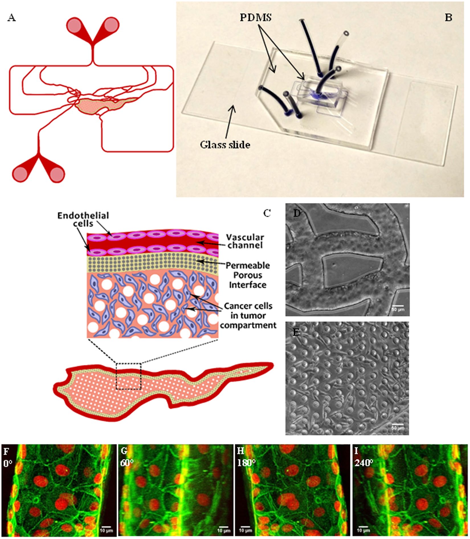
Schematic of the bMTM (A) with magnified view of the vascular compartment, vascular-tumor compartment interface and the tumor compartment (B). Optical image of the bMTM (C) with HBTAEC cultured in the vascular compartment (D) and MDA-MB-231 cultured in the tumor compartment (E). HBTAEC cultured under flow in the vascular compartment of bMTM form a complete lumen as shown with 3D reconstruction of confocal images of HBTAEC cultured in bMTM stained with f-actin (green) and Draq5 (red) after 4 days in culture maintained under flow of 0.05 μl/min (F–I); images are shown with a Y-axis rotation of 0, 60, 180 and 240 degrees in (F,G,H and I) respectively.
 Schematic of the bMTM (A) with magnified view of the vascular compartment, vascular-tumor compartment interface and the tumor compartment (B). Optical image of the bMTM (C) with HBTAEC cultured in the vascular compartment (D) and MDA-MB-231 cultured in the tumor compartment (E). HBTAEC cultured under flow in the vascular compartment of bMTM form a complete lumen as shown with 3D reconstruction of confocal images of HBTAEC cultured in bMTM stained with f-actin (green) and Draq5 (red) after 4 days in culture maintained under flow of 0.05 μl/min (F–I); images are shown with a Y-axis rotation of 0, 60, 180 and 240 degrees in (F,G,H and I) respectively.