The objective of our research is to develop a new type of ambient mass spectrometric technique combining non-resonant fs laser vaporization and electrospray ionization. The two methods combined allows for the vaporization of neutral sample molecules using the nonresonant fs laser and post-ionization using the electrospray. Using laser electrospray mass spectrometry (LEMS) we have successfully analyzed explosives, pharmaceuticals, large biomolecules and tissue.
Ambient Mass Spectrometry of Neutral Samples:
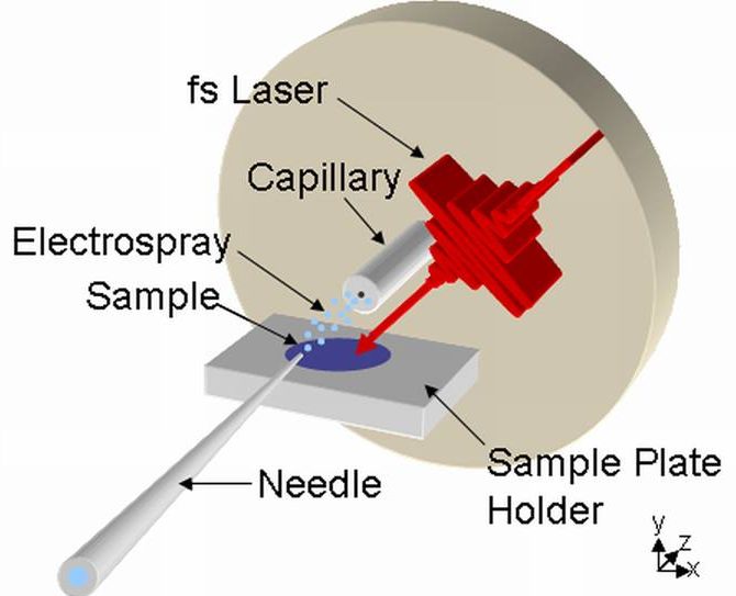
- LEMS combines non-resonant femtosecond laser vaporization and electrospray post-ionization.
- Neutral molecules are vaporized from a sample surface using a femtosecond laser.
- The neutral molecules are captured in the electrospray plume, where they undergo ionization.
- The ions travel to the inlet of the mass spectrometer for detection and mass analysis.
LEMS of Vitamin B12
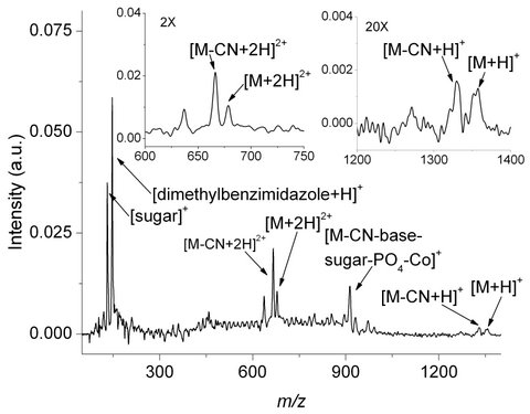
A 250 mL aliquot of 10-3 M vitamin B12 was spotted and dried on a glass slide and analyzed using LEMS. The acquired mass spectrum shows the protonated molecular ion at m/z 1356. The doubly charged ion is also observed demonstrating that electrospray ionization is the dominante mechanism. This is the first demonstration of matrix-free vaporization of neutral molecules at atmospheric conditions with a nonresonant femtosecond laser.
LEMS of Human Blood
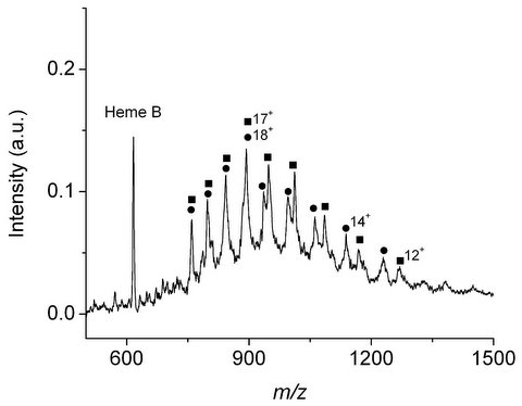
Human blood deposited on a metal slide was subjected to LEMS analysis. Multiple charging of hemoglobin subunits (a and b hemoglobin subunits are denoted by the squares and circles, respectively) indicates electrospray ionization is the dominant mechanism. In addition to the a and b hemoglobin subunits, the b heme is also present in the mass spectrum. We have demonstrated, using femtosecond laser pulses, that the vaporization of intact molecules up to 16,000 Da under ambient conditions is possible.
Mass Spectral Imaging using LEMS
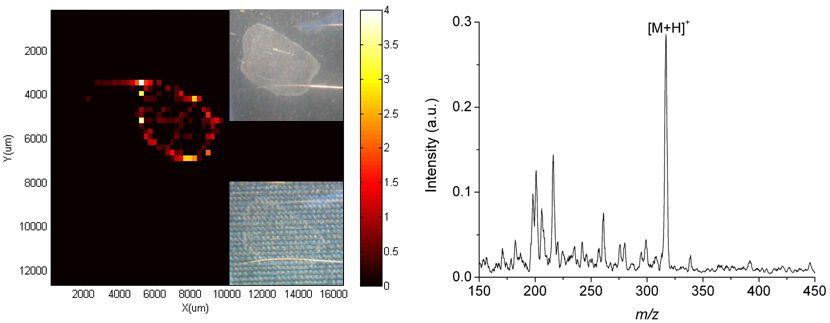
A 50 mL aliquot of 10-5 M oxycodone was spotted and dried on a stainless steel slide and analyzed using LEMS. A typical mass spectrum from red spots on the reconstructed image shows protonated adduct of oxycodone at m/z 316. No oxycodone is observed in the light gray spectrum which is consistent with the black areas on the reconstructed image. Inset pictures show before and after LEMS analysis of oxycodone.
LEMS Publications:
- Brady, J.J., Judge, E.J., and Levis, R.J., Mass spectrometry of intact neutral macromolecules using intense non-resonant femtosecond laser vaporization with electrospray post-ionization, Rapid Commun. Mass Spectrom., (2009) 23, 3151-3157.
- Judge, E.J., Brady, J.J., Dalton, D.R. and Levis, R.J., Analysis of pharmaceutical compounds from glass, fabric, steel and wood surfaces at atmospheric pressure using non-resonant femtosecond laser vaporization, electrospray mass spectrometry, Anal. Chem., (2010) 82, 3231-3238.
- Brady, J.J., Judge, E.J., and Levis, R.J., Identification of Explosives and Explosive Formulations Using Laser Electrospray Mass Spectrometry, Rapid Commun. Mass Spectrom., (2010) 24, 1659-1664.
- Judge, E.J., Brady, J.J., and Levis, R.J., Mass analysis of biological macromolecules at atmospheric pressure using nonresonant femtosecond laser vaporization and electrospray ionization, Anal. Chem., (2010) 82, 10203-10207.
- Brady, J.J., Judge, E.J., and Levis, R.J., Analysis of Amphiphilic Lipids and Hydrophobic Proteins Using Nonresonant Femtosecond Laser Vaporization with Electrospray Post-Ionization, J. Am. Soc. Mass Spectrom., (2011) 22, 762-772.
- Judge, E.J., Brady, J.J., Barbano, P.E., and Levis, R.J., Nonresonant Femtosecond Laser Vaporization with Electrospray Postionization for ex vivo Plant Tissue Typing Using Compressive Linear Classification, Anal. Chem., (2011) 83, 2145-2151.
- Brady, J.J., Judge, E.J., and Levis, R.J., Nonresonant Femtosecond Laser Vaporization of Aqueous Protein Preserves Folded Structure, P. Natl. Acad. Sci., (2011) 108, 12217-12222.